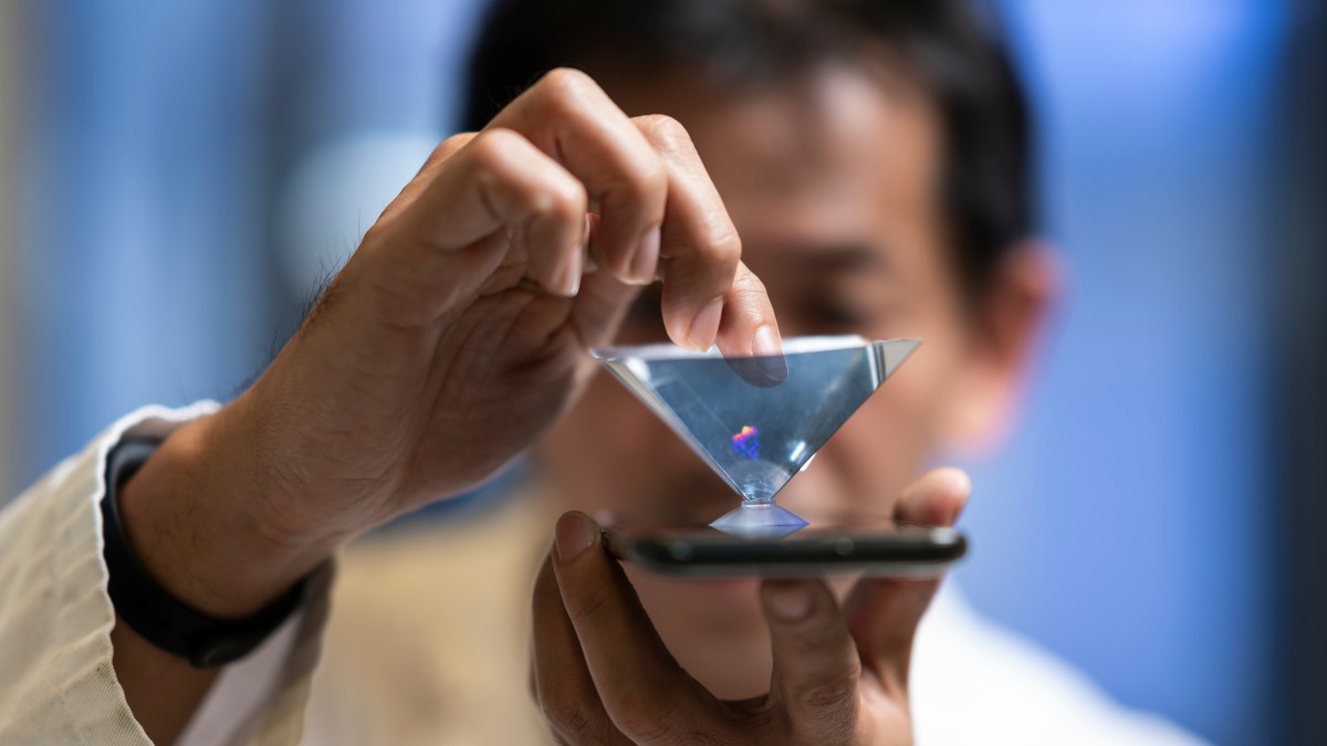
New portable technology that visualises clots forming in flowing blood in a 3D holographic livestream promises to dramatically improve screening and treatment of stroke, heart attack and coronavirus-induced lung failure.
Millions of people around the world die from heart attacks and strokes every year, and 2.5 million people have died due to coronavirus globally since the pandemic began.
In a world first, the biomedical invention from The Australian National University (ANU) can measure a blood clot’s “stickiness” and “optically weigh” it, within a thousand millionth of a gram, to assess a person’s disease risk.
ANU biomedical imaging scientist and research leader Dr Steve Lee said this technology advances his team’s breakthrough 2018 prototype diagnostic device in two critical ways. His multidisciplinary team has expertise in imaging sciences, medicine and biochemistry.
“We can now measure the stickiness of the blood clot down to a single platelet and we’ve dramatically reduced the size of our invention so that it can fit on a small desk or bench space in a hospital or another healthcare setting,” Dr Lee from the John Curtin School of Medical Research (JCSMR) said.
The new high-speed imaging technology, known as COSI (coherent optical scattering and interferometry), revealed in experiments how individual platelets “grip and walk” along a collagen fibre under blood flow.
“Platelets, which are a tenth of the size of a regular cell and are the major drivers of blood clot formation, move much like a circus performer walking along on a high wire,” Dr Lee said.
He said existing imaging tools are too slow to capture single platelet actions before they clump together within seconds of being activated.
“COSI has a very fast and high-resolution imaging process with no labeling, so it can capture the behaviour of individual platelets before they clump together,” Dr Lee said.
The breakthrough could be vital to study micro-blood clots in capillaries involved in lung failure related to COVID19, Dr Lee said.
Ms Yujie Zheng, the team’s lead PhD scholar, said seeing a platelet move in an orchestrated way within a developing blood clot, before suddenly freezing as she added a chemical inhibitor was a Eureka moment.
“That was a very exciting moment for us, because we could see these nanoscale events happening for the first time in a clot forming before our eyes,” Ms Zheng said.
Dr Lee’s team has worked closely with JCSMR’s National Platelet Research and Referral Centre led by Professor Elizabeth Gardiner. The collaboration has already received competitive research funding totalling $1.8 million.
“We have now moved beyond the proof of principle and are trialing COSI on a variety of patient samples with NPRC medical researchers with the aim to commercialise the technology within two years,” Dr Lee said.
The team’s work is published in the Biophysical Journal (Cell Press).


